-Blepharitis-

-Blepharitis is characterized by inflammation of the eyelids
-There is anterior and posterior blepharitis
-Anterior is characterized by inflammation of the base of the eyelashes. Less common than posterior
-Posterior is characterized by inflammation of the inner portion of the eyelid, at the level of the meibomian glands.
-can cause colonization of staphylococcal organisms
-can be associated with Rosacea and Seborrheic Dermatitis
-typically patients will have red eyes, a gritty sensation, excessive tearing, eyes may burn, swollen erythematous eyelids, crusting, and photophobia.
-lid hygiene is important for treatment
-warm compresses can be helpful
-topical antibiotics such azithromycin, erythromycin, or bacitracin may be helpful in reducing bacterial load of the lashes
-oral antibiotics such as tetracycline for severe cases of blepharitis
-Blowout Fracture-

-blowout fractures of the eye are fracture of the floor of the orbit
-typical mechanism is getting hit with a round object
-may causes entrapment of the inferior rectus muscle
-ischemia may cause loss of the muscle function and result in edema or hemorrhage of the muscle
-enophthalmos (the eyeball receded into the orbit) may develop when the globe is displaced posteriorly
-injury to the inferior orbital nerve may result from this causing sensory loss in that distribution
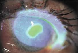
-Cataract-

-leading cause of blindness in the world
-it is an opacity in the lens of the eye that can cause total or partial blindness
-embryonic development and lifelong growth of the lens produce a complex layering of cells
-the lens does not shed its nonviable cells and this causes degenerative effects on its own cell structure leading some opacity problems
-risk factors for acquired cataracts are: age, smoking, alcohol, sunlight, metabolic syndrome, diabetes, statins, and long term use of high dose steroids
-no convincing evidence that vitamin supplementation decreases incidence
-only treatment is surgical correction
-Chalazion-

-Chalazion is an inflammatory lesion that develops when the meibomian tear gland becomes obstructed
-may first present as eyelid swelling and erythema, then evolve to a nodular rubbery lesion
-commonly seen in patients with blepharitis and rosacea
-antibiotics are not indicated
-frequent hot compresses are effective
-most of the time not painful or tender
-most of the time not painful or tender
-symptomatic patient can be referred to ophthalmology for incision and curettage or steroid injection
-Conjunctivitis-

-conjunctivitis is inflammation of the conjunctiva. Can be infectious or non infectious
-conjunctiva is usually transparent and gets red when inflamed
-infectious can be viral or bacterial
-non infectious can be allergic or non allergic
-bacterial typically caused by strep pneumonia, staphylococcus aureus, haemophilius influenzae, and moraxella catarrhalis
-can be caused by neisseria gonorrheae and chlamydia
-viral is usually adenovirus
-allergic conjunctivitis is caused by airborne allergens contacting the eye that cause mast cell degranulation
-itching is the cardinal symptom for allergic conjunctivitis
-can be non allergic from chronic dry eye
-contact lens wearer need to throw away contacts and irrigation solution and case
-bacterial conjunctivitis include erythromycin ointment or polytrim drops.
-Can also use sulfacetamide, azithromycin drops or bacitracin ointment
-Ciloxin needed for contact lens wears in bacterial conjunctivitis because of pseudomonas prominence
-for allergic conjunctivitis can use OTC decongestants, antihistamines, and patanol.
-students must receive topical therapy 24 hours before returning to school
-Corneal Abrasion-
 -usually result from trauma to the eye or from improper contact lens use
-diagnosis is made with slit lamp exam and fluoroscein dye exam
-treatment consists of topical antibiotics (drops or ointment) and oral pain medication
-most corneal abrasions heal within twenty-four hours after the accident
-patients present with photophobia, pain and foreign body sensation
-if a foreign body is detected can be removed with irrigation or swab after instillation of topical anesthetic
-superficial foreign bodies can be removed with a twenty five gauge needle or foreign body spud
-no patching of the eye
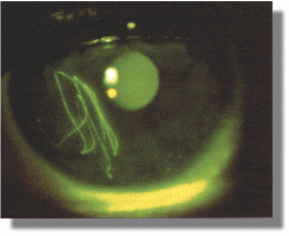
-Corneal Ulcer-
-corneal ulcers tend to be round on fluoroscein staining but are typically evident as a white spot on penlight or direct inspection
-diagnosis is made similar to a corneal abrasion with slit lamp and staining
-treatment is similar
-cover for herpes if there is any suspicion or if dendrites evident
-refer to ophthamology
-Dacrocystitis-
-dacrocystitis is caused by nasolacriminal duct being blocked
-can occur anywhere in the nasolacriminal duct system but mostly occurs at the distal end of the duct
-first line of treatment is massaging the tear duct
-probing may need to be done by an ophthalmologist
-acute dacrocystitis should be treated with antibiotics
-common organisms include alpha hemolytic strep, staph epidermis, and staph aureus
-coverage with clindamycin, doxycycline, or bactrim is helpful
-vancomycin is needed for more severe infection
-can lead to peri-orbital cellulitis
-Ectropion-

-Defined as an eversion of the eyelid away from the globe
-Frequency increases with age
-left untreated can cause dry eye and inflammation and damage the eye
-artificial tears should be used to treat dryness
-surgical treatment is directed at the etiology
-Entropion-
-diagnosis is made with slit lamp exam and fluoroscein dye exam
-treatment consists of topical antibiotics (drops or ointment) and oral pain medication
-most corneal abrasions heal within twenty-four hours after the accident
-patients present with photophobia, pain and foreign body sensation
-if a foreign body is detected can be removed with irrigation or swab after instillation of topical anesthetic
-superficial foreign bodies can be removed with a twenty five gauge needle or foreign body spud
-no patching of the eye
-Corneal Ulcer-
-corneal ulcers tend to be round on fluoroscein staining but are typically evident as a white spot on penlight or direct inspection
-diagnosis is made similar to a corneal abrasion with slit lamp and staining
-treatment is similar
-cover for herpes if there is any suspicion or if dendrites evident
-refer to ophthamology
-Dacrocystitis-
-dacrocystitis is caused by nasolacriminal duct being blocked
-can occur anywhere in the nasolacriminal duct system but mostly occurs at the distal end of the duct
-first line of treatment is massaging the tear duct
-probing may need to be done by an ophthalmologist
-acute dacrocystitis should be treated with antibiotics
-common organisms include alpha hemolytic strep, staph epidermis, and staph aureus
-coverage with clindamycin, doxycycline, or bactrim is helpful
-vancomycin is needed for more severe infection
-can lead to peri-orbital cellulitis
-Ectropion-

-Defined as an eversion of the eyelid away from the globe
-Frequency increases with age
-left untreated can cause dry eye and inflammation and damage the eye
-artificial tears should be used to treat dryness
-surgical treatment is directed at the etiology
-Entropion-

-Entropion is the turning in of an edge of an eyelid
-It usually is seen on the lower eyelid.
-trachoma an infection seen in the lower eyelid can cause
-trachoma can cause blindness but rarely seen in the US
-artificial tears should be used to help with dryness
-surgery is usually needed to correct condition
-It usually is seen on the lower eyelid.
-trachoma an infection seen in the lower eyelid can cause
-trachoma can cause blindness but rarely seen in the US
-artificial tears should be used to help with dryness
-surgery is usually needed to correct condition
-Foreign Body-

-Diagnosis is by gross examination, slit lamp exam and fluoroscein stain
-Slit lamp exam necessary to help determine the depth and position
-eyelids should be everted to look for residual foreign body
-attempt at foreign body removal can be made after topical anesthesia eye drops applied
-can be made with swab, 25 gauge needle, or eye-burr
-referral should be made to eye doctor if cannot be removed the same day or to ensure proper follow up
-no contact lenses until further instruction
-sit in a dark room, sunglasses to help with photophobia
-rust ring will need to be removed also with metal
-treated otherwise the same as corneal abrasions with eye drops or ointment
-Glaucoma-

-Glaucoma is a group of eye disorders that has elevated intra-ocular pressure (IOP)
-open angle glaucoma is an optic neuropathy that has a progressive peripheral visual loss followed by a central field loss, not always having increased IOP. Has cupping of the optic nerve
-acute closure glaucoma is characterized by closure of the anterior chamber angle and the aqueous humor cannot drain. This leads to increased IOP and damage to the optic nerve. It presents as a painful red eye that has to be treated within 24 hours to prevent blindness
-glaucoma can be secondary to uveitis, trauma, steroid therapy, vasoproliferative retinopathy, or ocular syndromes.
-glaucoma can be mixed
-the current evidence shows that lowering elevated IOP in glaucoma betters clinical outcomes
-screening only by IOP is inappropriate because pole with open angle glaucoma can have normal IOP
-IOP greater than 40 should dictate an emergency referral especially if symptomatic
-treatment with Xaltan drops
-Hordeolum-
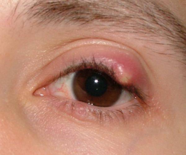
-a hordeolum (stye) is a painful red lump on the eyelid
-it happens when a gland on the edge of the eyelid gets inflamed
-most styes get better on their own after a few days
-a stye caused by an infection and is painful
-warm compresses are the mainstay of treatment
-also treatment with antibiotic eye ointment or drops
-Hyphema-
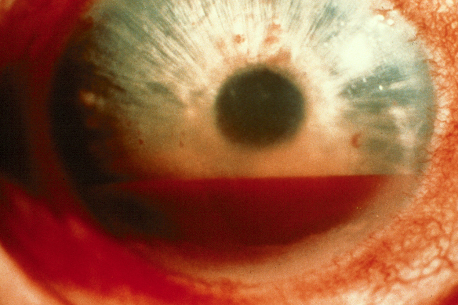
-a hyphema is blood in the anterior chamber of the eye. Usually caused by trauma
-can result in permanent vision loss
-early intervention by an ophthalmologist can decrease the likelihood of recurrent bleeding and avoid intraocular hypertension
-slit lamp exam necessary to look for micro hyphema
-emergent imaging is necessary if an open globe is suspected
-patients on anticoagulants or blood dyscrasias need labs including CBC and coagulation studies
-emergently limit bathroom privileges, keep head elevated to 30 degrees
-cycloplegia may help with pain control. Or oral analgesics
-patient likely will need surgical intervention to correct it
-Macular Degeneration-

-Macular degeneration is a degenerative disease of the central portion of the retina (macula) that results in central vision loss
-it is the leading cause of adult blindness and severe visual impairment
-there is wet (neuromuscular or exudative) and dry (atrophic) macular degeneration
-you can see sub retinal drusen deposits with dry macular degeneration
-large soft drusen spots or RPE pigmentary clumping increases with wet macular degeneration
-risk factors include age, smoking, family history, diet, and cardiovascular disease
-central vision loss is affected
-treatment of macular degeneration is targeted at type but can involve risk factor modification, antioxidant vitamins and zinc, and laser surgery
-Nystagmus-

-Nystagmus is a twitching of the eye
-the two major types of nystagmus are jerk and pendular nystagmus
-types of jerk nystagmus are downbeat, upbeat, horizontal, torsional, and mixed
-the direction named is the direction of the fast phase
-other nystagmus come out during certain conditions peripheral gaze and positional
-jerk nystagmus are the result of asymmetry in vestibular input in the central or peripheral nervous system
-pendular nystagmus has sinusoidal oscillation without fast phases
-pendular nystagmus may occur in any direction and can sometimes only be on one eye
-Differential diagnosis included structural lesion, metabolic derangement, infections, and intoxications
-four types of therapy include: medications, botulinum injections, prism lenses, and surgery
-therapy is targeted at specific type
-baclofen and neurontin good for specific forms of nystagmus
-surgery is reserved for certain types mainly congenital nystagmus
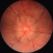
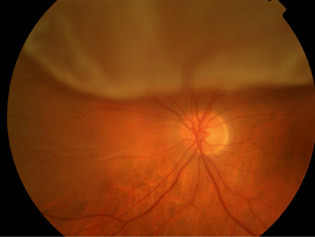
-Optic Neuritis-
-optic neuritis is an inflammatory, demyelinating condition that causes acute, usually monocular vision loss.
-optic neuritis is highly associated with multiple sclerosis
-it is the presenting symptom in 15-20 percent of MS cases
-usually monocular when presents, but can occur in both eyes 10 percent of the time
-vision loss develops over hours to days. Eye pain is usually present
-an afferent pupillary defect is present if the other eye is otherwise healthy
-the visual field defect is typically characterized with a central scotoma
-loss of color vision sometimes happens
-Papillitis with hyperemia and swelling of the optic disc and blurring of the disc margins is seen in optic neuritis
-optic neuritis can have infectious and non infectious etiologies also
-diagnosis is made by fundoscopic exam
-MRI can be helpful in the diagnosis and LP can by used for atypical cases
-treated with high dose steroids
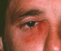
-Orbital Cellulitis-
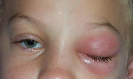
-orbital cellulitis is an infection that involves the fat and muscles around the orbit
-preseptal or periorbital cellulitis is an infection of the anterior portion of the eyelid
-infections do not effect the globe
-preseptal cellulitis is usually mild and almost never causes any serious complications
-orbital cellulitis can cause loss of vision or loss of life
-orbital cellulitis can cause ophthalmoplegia, pain with eye movements, and proptosis
-Imaging studies can be helpful to establish the diagnosis, CT or MRI
-orbital cellulitis is an uncommon complication of bacterial sinusitis
-other causes of orbital cellulitis are eye surgery, peri-bulbar anesthesia, orbital trauma with fracture or foreign body, dacrocystitis, dental infections, otitis media, or infected mucocele that erodes into the orbit
-fungal etiologies and TB are rare causes
-most common bacteria are streptococcus aureus and streptococci
-the infection can be complicated with an abscess, vision loss, cavernous sinus thrombosis, and brain abscess
-empiric treatment with vancomycin and rocephin or unasyn or zosyn
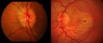
-Papilledema-
-papilledema is a swollen optic nerve secondary to an increased intracranial pressure
-cause of papilledema include: intracranial mass lesions, cerebral edema, increased CSF production (pseudotumor cerebri), decreased CSF reabsorption, obstructive hydrocephalus, obstruction of venous outflow (venous sinus thrombosis, and jugular vein compression)
-Papilledema is usually bilateral
-may have headache, nausea, vomiting, and blurry vision out of affected eye
-MRI of brain and Lumbar Puncture with opening pressure helpful for determining etiology
-Treatment is directed at the etiology. If pseudotumor cerebri is the cause diamox is helpful
-Pterygium-

-pterygium is a triangular wedge of conjunctival tissue that usually starts medially on the nasal conjunctiva and extends laterally onto the cornea
-classified a a corneal degenerative disorder
-may be a more proliferative condition with possible exacerbating factors of UV light, abnormal conjunctival tumor suppressor gene, presence of angiogenesis related factors, HPV infection, or abnormal HLA expression
-gets its name because looks like an insect wing
-usually occurs over months to years
-usually just observe unless causes visual impairment
-may induce an astigmatism (defect in the cornea)
-dryness can be treated with artificial tears
-surgery should be avoided for cosmetic reasons alone
-surgery should be done to correct a visual impairment or astigmatism that is induced
-Retinal Detachment-
-Retinal detachment happens when the multilayer retina separates from the underlying retinal pigment epithelium from the choroid
-this may occur passively due to accumulation of fluid between these two layers
-may also be due to vitreous traction of the retina, with diabetic traction retinal detachment
-separation of the retina results in ischemia and rapid progressive photoreceptor degeneration
-without treatment, most symptomatic retinal detachments involve the entire retina and will lead to loss of vision
-can be traumatic in etiology also
-risk factors include myopia, cataract surgery, PVD, ocular trauma, diabetes, and family history of retinal detachment
-patients typically complain of increasing number of floaters in one eye
-floaters may resemble a cobweb
-patients may notice a flash of light
-patients who present with a sudden onset of floaters, flashes of light, and monocular decreased visual acuity of field loss should be seen urgently by an ophthalmologist.
-diagnosis is made by fundoscopic exam
-treatment is dependent on type but is often corrective surgery
-Retinal Vascular Occlusion-

there are 3 main groups of retinal occlusion branch retinal vein occlusion, central retinal vein occlusion, and hemiretinal vein occlusion
-it is the second most common cause of vision loss from retinal vascular disease behind diabetic retinopathy
-risk factors include age, hypertension, diabetes, smoking, hypercoagulable states, glaucoma and retinal arteriolar abnormalities
-some patients are asymptomatic diagnosed on routine fundoscopic exam
-symptomatic patients may have scotoma or visual field defect with blurred or gray vision. If the macula is involved, they complain of blurred central vision
-patients who undergo neovasuclarization of the anterior chamber may acquire glaucoma
-eye exam should entail extra ocular movements, intraocular pressure, pupillary function, confrontation visual fields, slit lamp exams, visual acuity and dilated fundoscopic exam
-patients with branch and central retinal vascular occlusion should have retinal hemorrhage and edema, in addition to dilated retinal veins
-cotton wool spots are observed in approximately half of patients with central retinal vascular occlusion
-Fluorescein angiogram aids in the diagnosis
-Optical coherence tomography allows for high resolution cross sectioning of the retina
-in the absence of macular edema or neovascularization, there is no evidence that treatment improves outcomes and can be associated with sequela
-treatment when there is neovascularization and macular edema can involve laser photocoagulation, and medical therapy with vascular endothelial growth factor inhibitors or intravitreal glucocorticoids
-Retinopathy-

-diabetic retinopathy may cause vision loss by macular edema, hemorrhage from new vessels, retinal detachment, or neovascular glaucoma
-diabetic retinopathy has two types non proliferative (no new blood vessels) and proliferative (has new blood vessels)
-non proliferative retinopathy has nerve fiber layer infarcts (cotton wool spots), intra-retinal hemorrhages, hard exudates, and microvascular abnormalities
-proliferative retinopathy has neovascularization arising from the disc or retinal vessels, vitreous hemorrhages, fibrosis, and traction retinal detachment
-macular edema can occur at any stage of diabetic retinopathy
-the majority of patients who develop diabetic retinopathy have no symptoms until very late stages and can be too late for effective treatment
-the best treatment is prevention and control of diabetes and risk factor modification
-more rigorous control of blood pressure slows the rate of progression of diabetic retinopathy
-surgical intervention is helpful for certain stages and types of diabetic retinopathy
-another type of retinopathy, Retinopathy of Prematurity occurs in the retina of preterm infants with incomplete retinal vascularization.
-It is the most common cause of childhood blindness in the US
-the rate of retinopathy of prematurity increases with decreased gestational age
-risk factors include low birth weight, low gestational age, ventilation for greater than 7 days, surfactant therapy, high blood transfusion volume, hyperglycemia, fluctuations of blood gas measurements, and insulin therapy
-treatment consists of ablation of the peripheral avascular retina with laser therapy
-other forms of retinopathy include hypertensive retinopathy and atherosclerotic retinopathy caused by hypertension and atherosclerosis
-treatment is directed risk factor modification
-Strabismus-
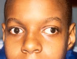
-strabismus is used to describe an anomaly of ocular alignment
-can occur in one or both eyes in any directs
-a latent strabismus is only present when the fixation is interrupted
-cranial nerve IV (trochlear nerve) innervates the superior oblique muscle, the lateral rectus muscle is innervated by the abducens nerve (cranial nerve VI) and the others by cranial nerve III (oculomotor)
-causes of strabismus include idiopathic, infantile esotropia, intermittent exotropia, and other congenital syndromes
-other secondary causes include retinoblastoma, optic nerve hypoplasia, macular scars, head trauma, cranial nerve palsies, orbital fracture, myasthenia gravis, and Graves Disease
-a trial of medical and physical therapy may precede surgical intervention in some cases of strabismus
-Slit lamp exam necessary to help determine the depth and position
-eyelids should be everted to look for residual foreign body
-attempt at foreign body removal can be made after topical anesthesia eye drops applied
-can be made with swab, 25 gauge needle, or eye-burr
-referral should be made to eye doctor if cannot be removed the same day or to ensure proper follow up
-no contact lenses until further instruction
-sit in a dark room, sunglasses to help with photophobia
-rust ring will need to be removed also with metal
-treated otherwise the same as corneal abrasions with eye drops or ointment
-Glaucoma-
-Glaucoma is a group of eye disorders that has elevated intra-ocular pressure (IOP)
-open angle glaucoma is an optic neuropathy that has a progressive peripheral visual loss followed by a central field loss, not always having increased IOP. Has cupping of the optic nerve
-acute closure glaucoma is characterized by closure of the anterior chamber angle and the aqueous humor cannot drain. This leads to increased IOP and damage to the optic nerve. It presents as a painful red eye that has to be treated within 24 hours to prevent blindness
-glaucoma can be secondary to uveitis, trauma, steroid therapy, vasoproliferative retinopathy, or ocular syndromes.
-glaucoma can be mixed
-the current evidence shows that lowering elevated IOP in glaucoma betters clinical outcomes
-screening only by IOP is inappropriate because pole with open angle glaucoma can have normal IOP
-IOP greater than 40 should dictate an emergency referral especially if symptomatic
-treatment with Xaltan drops
-Hordeolum-
-a hordeolum (stye) is a painful red lump on the eyelid
-it happens when a gland on the edge of the eyelid gets inflamed
-most styes get better on their own after a few days
-a stye caused by an infection and is painful
-warm compresses are the mainstay of treatment
-also treatment with antibiotic eye ointment or drops
-Hyphema-
-a hyphema is blood in the anterior chamber of the eye. Usually caused by trauma
-can result in permanent vision loss
-early intervention by an ophthalmologist can decrease the likelihood of recurrent bleeding and avoid intraocular hypertension
-slit lamp exam necessary to look for micro hyphema
-emergent imaging is necessary if an open globe is suspected
-patients on anticoagulants or blood dyscrasias need labs including CBC and coagulation studies
-emergently limit bathroom privileges, keep head elevated to 30 degrees
-cycloplegia may help with pain control. Or oral analgesics
-patient likely will need surgical intervention to correct it
-Macular Degeneration-
-Macular degeneration is a degenerative disease of the central portion of the retina (macula) that results in central vision loss
-it is the leading cause of adult blindness and severe visual impairment
-there is wet (neuromuscular or exudative) and dry (atrophic) macular degeneration
-you can see sub retinal drusen deposits with dry macular degeneration
-large soft drusen spots or RPE pigmentary clumping increases with wet macular degeneration
-risk factors include age, smoking, family history, diet, and cardiovascular disease
-central vision loss is affected
-treatment of macular degeneration is targeted at type but can involve risk factor modification, antioxidant vitamins and zinc, and laser surgery
-Nystagmus-
-Nystagmus is a twitching of the eye
-the two major types of nystagmus are jerk and pendular nystagmus
-types of jerk nystagmus are downbeat, upbeat, horizontal, torsional, and mixed
-the direction named is the direction of the fast phase
-other nystagmus come out during certain conditions peripheral gaze and positional
-jerk nystagmus are the result of asymmetry in vestibular input in the central or peripheral nervous system
-pendular nystagmus has sinusoidal oscillation without fast phases
-pendular nystagmus may occur in any direction and can sometimes only be on one eye
-Differential diagnosis included structural lesion, metabolic derangement, infections, and intoxications
-four types of therapy include: medications, botulinum injections, prism lenses, and surgery
-therapy is targeted at specific type
-baclofen and neurontin good for specific forms of nystagmus
-surgery is reserved for certain types mainly congenital nystagmus
-Optic Neuritis-
-optic neuritis is an inflammatory, demyelinating condition that causes acute, usually monocular vision loss.
-optic neuritis is highly associated with multiple sclerosis
-it is the presenting symptom in 15-20 percent of MS cases
-usually monocular when presents, but can occur in both eyes 10 percent of the time
-vision loss develops over hours to days. Eye pain is usually present
-an afferent pupillary defect is present if the other eye is otherwise healthy
-the visual field defect is typically characterized with a central scotoma
-loss of color vision sometimes happens
-Papillitis with hyperemia and swelling of the optic disc and blurring of the disc margins is seen in optic neuritis
-optic neuritis can have infectious and non infectious etiologies also
-diagnosis is made by fundoscopic exam
-MRI can be helpful in the diagnosis and LP can by used for atypical cases
-treated with high dose steroids
-Orbital Cellulitis-
-orbital cellulitis is an infection that involves the fat and muscles around the orbit
-preseptal or periorbital cellulitis is an infection of the anterior portion of the eyelid
-infections do not effect the globe
-preseptal cellulitis is usually mild and almost never causes any serious complications
-orbital cellulitis can cause loss of vision or loss of life
-orbital cellulitis can cause ophthalmoplegia, pain with eye movements, and proptosis
-Imaging studies can be helpful to establish the diagnosis, CT or MRI
-orbital cellulitis is an uncommon complication of bacterial sinusitis
-other causes of orbital cellulitis are eye surgery, peri-bulbar anesthesia, orbital trauma with fracture or foreign body, dacrocystitis, dental infections, otitis media, or infected mucocele that erodes into the orbit
-fungal etiologies and TB are rare causes
-most common bacteria are streptococcus aureus and streptococci
-the infection can be complicated with an abscess, vision loss, cavernous sinus thrombosis, and brain abscess
-empiric treatment with vancomycin and rocephin or unasyn or zosyn
-Papilledema-
-papilledema is a swollen optic nerve secondary to an increased intracranial pressure
-cause of papilledema include: intracranial mass lesions, cerebral edema, increased CSF production (pseudotumor cerebri), decreased CSF reabsorption, obstructive hydrocephalus, obstruction of venous outflow (venous sinus thrombosis, and jugular vein compression)
-Papilledema is usually bilateral
-may have headache, nausea, vomiting, and blurry vision out of affected eye
-MRI of brain and Lumbar Puncture with opening pressure helpful for determining etiology
-Treatment is directed at the etiology. If pseudotumor cerebri is the cause diamox is helpful
-Pterygium-
-pterygium is a triangular wedge of conjunctival tissue that usually starts medially on the nasal conjunctiva and extends laterally onto the cornea
-classified a a corneal degenerative disorder
-may be a more proliferative condition with possible exacerbating factors of UV light, abnormal conjunctival tumor suppressor gene, presence of angiogenesis related factors, HPV infection, or abnormal HLA expression
-gets its name because looks like an insect wing
-usually occurs over months to years
-usually just observe unless causes visual impairment
-may induce an astigmatism (defect in the cornea)
-dryness can be treated with artificial tears
-surgery should be avoided for cosmetic reasons alone
-surgery should be done to correct a visual impairment or astigmatism that is induced
-Retinal Detachment-
-Retinal detachment happens when the multilayer retina separates from the underlying retinal pigment epithelium from the choroid
-this may occur passively due to accumulation of fluid between these two layers
-may also be due to vitreous traction of the retina, with diabetic traction retinal detachment
-separation of the retina results in ischemia and rapid progressive photoreceptor degeneration
-without treatment, most symptomatic retinal detachments involve the entire retina and will lead to loss of vision
-can be traumatic in etiology also
-risk factors include myopia, cataract surgery, PVD, ocular trauma, diabetes, and family history of retinal detachment
-patients typically complain of increasing number of floaters in one eye
-floaters may resemble a cobweb
-patients may notice a flash of light
-patients who present with a sudden onset of floaters, flashes of light, and monocular decreased visual acuity of field loss should be seen urgently by an ophthalmologist.
-diagnosis is made by fundoscopic exam
-treatment is dependent on type but is often corrective surgery
-Retinal Vascular Occlusion-
there are 3 main groups of retinal occlusion branch retinal vein occlusion, central retinal vein occlusion, and hemiretinal vein occlusion
-it is the second most common cause of vision loss from retinal vascular disease behind diabetic retinopathy
-risk factors include age, hypertension, diabetes, smoking, hypercoagulable states, glaucoma and retinal arteriolar abnormalities
-some patients are asymptomatic diagnosed on routine fundoscopic exam
-symptomatic patients may have scotoma or visual field defect with blurred or gray vision. If the macula is involved, they complain of blurred central vision
-patients who undergo neovasuclarization of the anterior chamber may acquire glaucoma
-eye exam should entail extra ocular movements, intraocular pressure, pupillary function, confrontation visual fields, slit lamp exams, visual acuity and dilated fundoscopic exam
-patients with branch and central retinal vascular occlusion should have retinal hemorrhage and edema, in addition to dilated retinal veins
-cotton wool spots are observed in approximately half of patients with central retinal vascular occlusion
-Fluorescein angiogram aids in the diagnosis
-Optical coherence tomography allows for high resolution cross sectioning of the retina
-in the absence of macular edema or neovascularization, there is no evidence that treatment improves outcomes and can be associated with sequela
-treatment when there is neovascularization and macular edema can involve laser photocoagulation, and medical therapy with vascular endothelial growth factor inhibitors or intravitreal glucocorticoids
-Retinopathy-
-diabetic retinopathy may cause vision loss by macular edema, hemorrhage from new vessels, retinal detachment, or neovascular glaucoma
-diabetic retinopathy has two types non proliferative (no new blood vessels) and proliferative (has new blood vessels)
-non proliferative retinopathy has nerve fiber layer infarcts (cotton wool spots), intra-retinal hemorrhages, hard exudates, and microvascular abnormalities
-proliferative retinopathy has neovascularization arising from the disc or retinal vessels, vitreous hemorrhages, fibrosis, and traction retinal detachment
-macular edema can occur at any stage of diabetic retinopathy
-the majority of patients who develop diabetic retinopathy have no symptoms until very late stages and can be too late for effective treatment
-the best treatment is prevention and control of diabetes and risk factor modification
-more rigorous control of blood pressure slows the rate of progression of diabetic retinopathy
-surgical intervention is helpful for certain stages and types of diabetic retinopathy
-another type of retinopathy, Retinopathy of Prematurity occurs in the retina of preterm infants with incomplete retinal vascularization.
-It is the most common cause of childhood blindness in the US
-the rate of retinopathy of prematurity increases with decreased gestational age
-risk factors include low birth weight, low gestational age, ventilation for greater than 7 days, surfactant therapy, high blood transfusion volume, hyperglycemia, fluctuations of blood gas measurements, and insulin therapy
-treatment consists of ablation of the peripheral avascular retina with laser therapy
-other forms of retinopathy include hypertensive retinopathy and atherosclerotic retinopathy caused by hypertension and atherosclerosis
-treatment is directed risk factor modification
-Strabismus-
-strabismus is used to describe an anomaly of ocular alignment
-can occur in one or both eyes in any directs
-a latent strabismus is only present when the fixation is interrupted
-cranial nerve IV (trochlear nerve) innervates the superior oblique muscle, the lateral rectus muscle is innervated by the abducens nerve (cranial nerve VI) and the others by cranial nerve III (oculomotor)
-causes of strabismus include idiopathic, infantile esotropia, intermittent exotropia, and other congenital syndromes
-other secondary causes include retinoblastoma, optic nerve hypoplasia, macular scars, head trauma, cranial nerve palsies, orbital fracture, myasthenia gravis, and Graves Disease
-a trial of medical and physical therapy may precede surgical intervention in some cases of strabismus
Here are a few herbal remedies for Retinal Vein Occlusion Herbal Treatment that are Bilberry, Gotu kola, Arjuna, Beta Carotene, Zinc, etc.
ReplyDeleteThe information you have posted is very useful. The sites you have referred was good. Thanks for sharing.. Lasik Eye Surgery Michigan
ReplyDeleteI havent any word to appreciate this post.....Really i am impressed from this post....the person who create this post it was a great human..thanks for shared this with us. how much does eye lift surgery cost
ReplyDeleteHi! Thanks for the great information you havr provided! You have touched on crucuial points! yaldoeyecenter.com
ReplyDeleteYou know your projects stand out of the herd. There is something special about them. It seems to me all of them are really brilliant! Yaldo Eye Center
ReplyDeleteAm Richard, I am here to testify about a great herbalist man who cured my wife of breast cancer. His name is Dr Imoloa. My wife went through this pain for 3 years, i almost spent all i had, until i saw some testimonies online on how Dr. Imoloa cure them from their diseases, immediately i contacted him through. then he told me the necessary things to do before he will send the herbal medicine. Wish he did through DHL courier service, And he instructed us on how to apply or drink the medicine for good two weeks. and to greatest surprise before the upper third week my wife was relief from all the pains, Believe me, that was how my wife was cured from breast cancer by this great man. He also have powerful herbal medicine to cure diseases like: Alzheimer's disease, parkinson's disease, vaginal cancer, epilepsy Anxiety Disorders, Autoimmune Disease, Back Pain, Back Sprain, Bipolar Disorder, Brain Tumor, Malignant, Bruxism, Bulimia, Cervical Disc Disease, Cardiovascular Disease, Neoplasms , chronic respiratory disease, mental and behavioral disorder, Cystic Fibrosis, Hypertension, Diabetes, Asthma, Autoimmune inflammatory media arthritis ed. chronic kidney disease, inflammatory joint disease, impotence, alcohol spectrum feta, dysthymic disorder, eczema, tuberculosis, chronic fatigue syndrome, constipation, inflammatory bowel disease, lupus disease, mouth ulcer, mouth cancer, body pain, fever, hepatitis ABC, syphilis, diarrhea, HIV / AIDS, Huntington's disease, back acne, chronic kidney failure, addison's disease, chronic pain, Crohn's pain, cystic fibrosis, fibromyalgia, inflammatory Bowel disease, fungal nail disease, Lyme disease, Celia disease, Lymphoma, Major depression, Malignant melanoma, Mania, Melorheostosis, Meniere's disease, Mucopolysaccharidosis, Multiple sclerosis, Muscular dystrophy, Rheumatoid arthritis. You can reach him Email Via drimolaherbalmademedicine@gmail.com / whatsapp +2347081986098 Website/ www.drimolaherbalmademedicine.wordpress.com
ReplyDeleteColorectal cancer, cancer of the large intestine, is the fourth most common cancer in North America. Many cases of colorectal cancer are associated with low levels of physical activity and with diets that are low in fruits and vegetables. Individuals with a family history of the disease have a higher risk. I crumble with this disease for 5 years also with a lot of scaring thought in my head because i was just waiting for death every day of my life until My Son came to me in the hospital explaining to me that he has find a herbal healer from Nigeria to cure my Colo-Rectal Cancer,I was so shocked with the ideal also i was excited inside of me.My son asked me to let us give him a try because we have really heard a lot of scammer pretending to cure all sort of diseases with herbal medicine and some of them never get a positive result at the end of it all but we was very confident on this herbal doctor,like i said we give him a try and he sent me a herbal medicine to drink for three weeks, Sincerely I'm telling you today I' alive and healthy no more laying on sick bed,No more Colo-Rectal Cancer.I'm sharing this testimony on here for people who are sick to contact this Wonderful man,His name is Dr Itua.And His contact Whatsapp_+2348149277967____Email_drituaherbalcenter@gmail.com.
ReplyDeleteHe can cure those diseases like:
Bladder cancer
Breast cancer
Colorectal cancer
Kidney cancer
Leukemia
Lung cancer
Non-Hodgkin lymphoma
Prostate cancer
Skin cancer
Uterine cancer
Hiv/Aids
Stroke
Herpes
Hepatitis
Love Spell
Diabetes
Most prostate cancers are adenocarcinomas, cancers that arise in glandular cells of the prostate’s epithelial tissue. Prostate cancers usually progress slowly and produce no symptoms in the initial stages. Eventually, the tumor may enlarge like mine too, the prostate gland, pressing on the urethra and causing painful or frequent urination and blood in the urine. So I was so uncomfortable with this prostate cancer diseases then I decided to do online search on how to cure cancer because I well have read a lot about herbal medicine, I came across a lot of testimony how Dr Itua cure HIV/herpes then Cancer was listed below the comment.with courage I contacted Dr Itua and he sent me his herbal medicine through Courier service then I was asked to pick it up at my post office which i quickly did. I contacted Dr Itua that I have received my herbal medicine so he instructed me on how to drink it for three weeks and that is how Dr Itua Herbal Medicine cures my prostate Cancer, The treatment takes three weeks and I was cured completely. Dr Itua is a god sent and I thank him every day of my life. Contact him now On: Email:drituaherbalcenter@gmail.com/ Whatsapp:+2348149277967.
ReplyDeleteHe listed that he can as well cure the following diseases below.... Cerebral Amides. Lung Cancer,Brain cancer,Esophageal cancer,Gallbladder cancer,Gestational trophoblastic disease,Head and neck cancer,Hodgkin lymphoma Intestinal cancer,Kidney cancer,Leukemia,Liver cancer,Melanoma,Mesothelioma,Multiple myeloma,Neuroendocrine tumors,Hodgkin lymphoma,Oral cancer,Ovarian cancer,Sinus cancer,Soft tissue sarcoma,Spinal cancer,Stomach cancer,Meniere's disease , Testicular cancer,Throat cancer,Thyroid Cancer,Uterine cancer,Vaginal cancer,Vulvar cancer. Alzheimer's disease,Autism,measles, tetanus, whooping cough, tuberculosis, polio and diphtheria Adrenocortical carcinoma. Alma, Uterine Cancer, Breast Cancer, Allergic diseases. Kidney cancer, Love Spell, Glaucoma., Cataracts,Macular degeneration,Cardiovascular disease,Lung disease.Enlarged prostate,Osteoporosis.Generalized dermatitis,Alzheimer's disease,Brain Tumor,Lupus,Endometrial Cancer, cerebrovascular diseases
Dementia.Colo rectal cancer, Lottery Spell, Bladder Cancer, Skin Cancer,Ovarian Cancer,Pancreatic Cancer, HIV /Aids,Brain Tumor, Herpes, Non-Hodgkin lymphoma, Inflammatory bowel disease, Copd, Diabetes, Hepatitis