-Acute Pharyngitis-

-Acute pharyngitis is one of the most common conditions encountered in the office practice
-Group A Streptococcus (GAS) makes up about 5-15 percent of the adults presenting with pharyngitis
-Etiologies include GAS, mononucleosis, CMV, HSV, and many other viruses including influenza
-Non Group A Streptococcus a groups C and G cause pharyngitis but do not cause rheumatic fever
-Diphtheria, Neisseria Gonorrheae, Chlamydia, Mycoplasma, and Tularemia can cause also
-Symptoms include sore throat, worse with swallowing, headache, malaise, and anterior neck pain form lymphadenopathy
-Exam may reveal pharyngeal erythema, tonsillar hypertrophy, and purulent exudates. Palate petechiae may be present with lymphadenopathy
-may have hepatosplenomegaly with mono
-Centor Criteria-tonsilar exudate, tender anterior cervical lymphadenopathy, fever, and absence of cough
-The presence of 3 or more Centor Criteria a rapid antigen detection test should be performed (Rapid Strep)
-If there is two or less of the Centor Criteria neither throat culture or rapid strep is necessary
-Other dangerous infections causing sore throat include epiglottis, peri-tonsilar abscess, submandibular space infection, retropharyngeal space infections and primary HIV
-Best to use tylenol or NSAIDS for systemic pain control
-Can use topical lozenges or sprays
-Use of glucocorticoids is controversial
-Penicillin VK, Amoxicillin or Bicllin CR times one dose treatment of choice
-Can use cephalosporins or macrolides for alternative therapy
-Augmentin for beta lactam resistance
-Aphthous Ulcers-
-aphthous ulcers are also known as canker sores
-they are painful lesions that appear as localized, shallow, round, ulcers with a grayish base
-local cell mediated immunity may be important in pathogenesis
-factors that predispose include trauma, hormonal factors, drug sensitivity, food sensitivity, immunodeficiency, and emotional stress
-also seen in patients with celiac disease or inflammatory bowel disease
-Vitamin and mineral deficiency have been implicated in the pathogenesis
-Treatment includes topical steroids and topical analgesics (magic mouth wash)
-Diseases of the Teeth and Gums-

-Periodontal disease is disease that effect the gingiva, cementum, periodontal ligament and the alveolar bone
-Periodontitis is inflammation of the gingiva and the alveolar bone
-Gingivitis dose not affect the alveolar bone. It is characterized by inflammation land gingival redness and swelling. Bleeds easy
-Gingivitis can be provoked by drug or pregnancy.
-Scurvy is a gingival disease provoked by Vitamin C Deficiency
-Necrotizing ulcerative gingivitis is called trench mouth. It can cause systemic symptoms, pain, ulcerative necrotic gingiva. It is associated immune disorders and malnutrition
-Dental infections can cause hematogenous disseminations of the infection and cause to seed native or prosthetic heart valves, joints or other devices
-Dry socket is osteomyelitis of the alveolar bone that can happen after dental extractions
-Dental caries come as a complication of dental decay. Treated with filling restoration and prevention
-Pulpitis is inflammation of dental pulp from dental decay presents with severe dental pain and temperature sensitivity
-Antibiotics help with local spread of infection and prevent hematogenous dissemination.
-It is also recommended for patients with prosthetic heart valves and artificial devices that get antibiotic prophylaxis prior to any procedure
-Usually penicillin VK is recommended or clindamycin
-Epiglottitis-

-Epiglottis is inflammation of the epiglottis and adjacent supraglottic structures
-Can lead to life threatening airway obstruction
-It results from direct spread of the epithelial layer of the bacteria
-Most common cause is Haemophilus Influenzae type B (HIB)
-Can be caused by many other bacteria or even viruses less likely
-The incidence of epiglottis has decreased with the HIB vaccine
-can come from noninfectious causes such as trauma, thermal injury or foreign body ingestion
-can come from fungal causes especially in immunocomprimised hosts
-presents with an abrupt onset of dysphagia, drooling, and respiratory distress. Also see fever and sore throat
-direct visualization confirms the diagnosis
-on soft tissue neck should see "thumb sign"
-should try to minimize agitation for exam if high index of suspicion. Have patient intubated in the OR with anesthesia
-Airway maintenance is the most important feature in the treatment of Epiglottis
-Intubation for 2-3 days is usually necessary before the patient can be safely extubated
-the role of glucocorticoids are controversial
-Empiric antibiotic therapy should include ceftriaxone and an anti-staph agent clindamycin or vancomycin
-racemic epinephrine may provide some temporary relief
-Laryngitis-
-Acute laryngitis is a self limited inflammatory condition lasting less than 3 weeks usually associated with a upper respiratory infection or voice strain
-The etiology is usually viral with acute laryngitis
-Strep Pneumonia, H. Influenzae, and M. Catarrhalis have been isolated in patients with acute laryngitis
-Acute laryngitis from a URI usually has sore throat, hoarse voice, and runny nose.
-Chronic laryngitis it typically associated with one or more chronic irritants, that cause inflammation
-Oral Candidiasis-

-Also known as thrush
-It is a common infection in infants. It occurs in adults who wear dentures, treated with antibiotics, radiation therapy, or chemotherapy
-Oral candidia also is seen in those in immunodeficient stats such as HIV
-Can occur with patients taking oral glucocorticoids
-Usual pathogen is candidia alblicans
-Pseudomembrane type or oral candidia is most common and is white plaques on buccal mucosa, palate, and tongue
-The other form is atrophic form also known as denture stomatitis. Cotton mouth, loss of taste, and pain with eating
-Treatment involves nystatin swish and spit for uncomplicated oral thrush
-Oral Herpes Simplex-

-the herpes simplex virus is spread by direct contact with the epidermis and eventually the sensory and autonomic nerve endings
-the lesions are painful and can last for 10-14 days
-the lesions present as grouped vesicles on an erythematous base
-these are also known as cold sores
-treatment is Acyclovir 200 mg PO five times a day or 400 mg TID
-you can also use Famciclovir 500 mg TID or Valacyclovir 1000 mg BID
-Oral Leukoplakia-

-Oral leukoplakia is a precancerous lesion that is white patches or plaques of the oral mucosa
-It is hyperplasia of squamous epithelium
-It is associated with human papilloma virus (HPV)
-One to twenty percent of these lesions will progress to carcinoma within 10 years
-Smokeless tobacco is a major risk factor
-Oral hairy leukoplakia occurs in HIV patients is not premalignant. It is caused by the Epstein Barr Virus
-These lesions need biopsied and monitored frequently
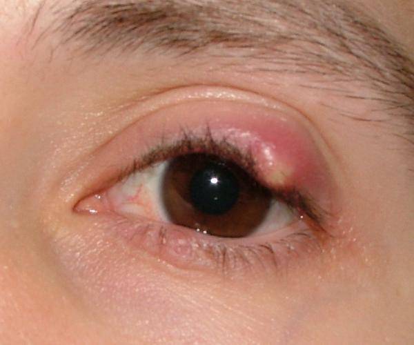
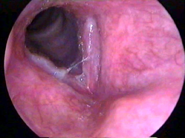
-Peritonsillar Abscess-

-Peritonsillar Abscess is a collection of pus between the capsule of the palatine tonsil and the pharyngeal muscles
-Peritonsillar infection is preceded by tonsillitis and progresses to cellulitis to phlegmon to abscess.
-It can also occur without preceding infection
-Peritonsillar abscess can compromise the upper airway and surrounding structures
-Peritonsillar abscess usually present with sore throat, fever, and a hot potato voice. Pooling of saliva or drooling may be present.
-Trismus may be present
-If there is these is a concern of possible epiglottis or the diagnosis of peritonsillar abscess is not definitive, imaging of the neck must be accomplished. CT scan with IV contrast of soft tissue neck
-Empiric antibiotics for Group A Strep and Staph aureus and respiratory anaerobes should be accomplished
-Unasyn or Clindamycin is appropriate antibiotic coverage
-If there is no response to therapy or there is airway issues vancomycin should be used also in addition to above antibiotics
-Ultimate treatment for peritonsillar abscess is incision and drainage
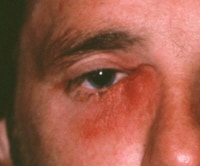
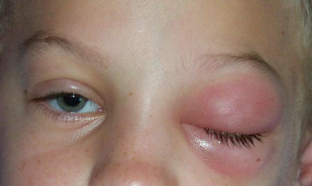
-Parotitis-

-Bacterial Parotitis is usually caused by Staph Aureus and mixed oral aerobes and anaerobes
-Can occur in the presences of dehydration and poor oral hygiene
-May occur from salivary stasis and retrograde seeding of the Stensen's Duct of the parotid gland with oral flora
-Stensen's duct may also be obstructed with a salivary stone or tumor
-On physical exam it presents as a sudden onset of firm erythematous swelling of the pre and posterior auricular area that extends to the angle of the mandible
-Staphylococcus aureus is the most isolate bacteria
-Imaging studies such as CT scan of soft tissue neck with IV contrast or Ultrasound is helpful to determine if abscess or stone is present
-Treatment is with IV antibiotics. Naficillin and Metronidazole recommended for immunocompetent patients.
-If patient is immunocompromised, vancomycin plus cefepime or imipenem is recommended
-Silaadenitis-

-typically presents with pain, swelling and erythema in the area of the gland
-there may be pus draining from the affected duct
-may be caused by salivary duct stone
-usually resolves within 7-10 days when treated with antibiotics
-if does not improve may develop an abscess and need to do a CT scan with IV contrast of the soft tissue neck
-dicloxacillin or cephalexin is ideal treatment for staph coverage
-Benign and Malignant Neoplasms of Oral Cavity-

-Squamous Cell Carcinoma of the mouth is associated with ulcers or masses that do not heal with dental changes or poorly fitted dentures
-Tongue and lip cancers present as ulcerative lesions usually painful
-Persistent plaques, ulcers, or erosions should be biopsied
-Melanoma-should be considered on oral pigmented lesions that have irregular borders and asymmetry or increasing diameter. Surgery is treatment of choice
-Amalgam Tattoos-are blue black macules seen in the gingiva near dental fillings. Benign lesions.
-Fordyce Spots-benign tumors of sebaceous gland etiology. These are isolated white to yellow papules prominent on the vermillion and mucosal border
-Mucoceles-fluid filled cavities with mucous glands lining of the epithelium. These are typically seen after mild oral trauma or disruption of the salivary duct. Rupture can lead to complete resolution.